![]() Figure 8 of
Zhu, Mol Vis 2002;
8:462-471.
Figure 8 of
Zhu, Mol Vis 2002;
8:462-471.
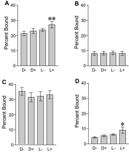
Figure 8. Phosphorylation- and light-dependent binding of mCARFL to chicken OS membranes
E19 chicken OS were phosphorylated for 30 min in the light with recombinant GRK1 and cold ATP (phosphorylated), along with a control containing no ATP or GRK (unphosphorylated). After regeneration of the opsins, 35S-labeled mCARFL (A), mCARΔE14 (B), mCAR clone 12 (C) and mSAG (D), were added to the phosphorylated and unphosphorylated OS in the dark and allowed to bind to the membranes in the light or dark for 10 min at 37 °C. After washing, the membranes were solubilized in SDS sample buffer, and electrophoresed next to a lane containing the total amount of arrestin added to each binding reaction. The gels were dried and subjected to phosphorimager detection of 35S products. D-, dark, unphosphorylated; D+, dark, phosphorylated; L-, light, unphosphorylated; L+, light, phosphorylated. p<0.05 (L+ versus D-); p<0.01 (L+ versus D-).