![]() Figure 7 of
Zhu, Mol Vis 2002;
8:462-471.
Figure 7 of
Zhu, Mol Vis 2002;
8:462-471.
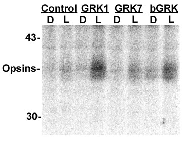
Figure 7. Phosphorylation of chicken OS membranes by GRKs
E19 chicken eyes were dark-adapted for 2 h, and the retinas were dissected under infrared light and shaken in 40% sucrose buffer to float the OS, which were then diluted and pelleted. The OS were phosphorylated in the dark (D) or light (L) for 10 min at room temperature in the presence of 8 μCi [γ-32P] ATP (6000 Ci/mmol) with no exogenous GRK (control) or 5 μl of exogenous recombinant GRK1, GRK7, or native bovine GRK (bGRK). The kinase reaction was stopped with SDS sample buffer, and the 32P labeled proteins were electrophoresed and detected in the dried gel with a phosphoimager. Molecular weight markers are shown on the left.