![]() Figure 4 of
Zhu, Mol Vis 2002;
8:462-471.
Figure 4 of
Zhu, Mol Vis 2002;
8:462-471.
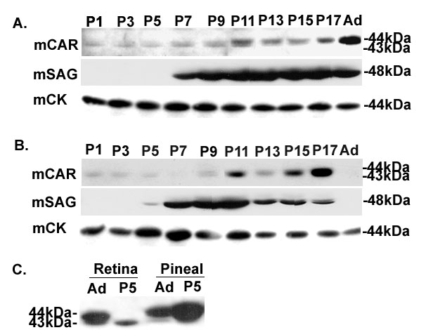
Figure 4. Developmental Study of mCAR expression
C57 (A) and rd/rd mice (B) were killed at P1, 3, 5, etc. and adult (Ad), and retinas were dissected. Fifty micrograms of soluble retinal proteins were applied to an 11.5% SDS-PAGE and processed for immunoblot analysis with primary antibodies LUMIJ for mCAR, C10C10 for rod arrestin (mSAG), and polyclonal antibody 1948 for creatine kinase (mCK) sequentially. Molecular weights of proteins are indicated on the right. (C) The expression pattern of mCAR in normal adult and P5 mouse retina and the pineal gland. Soluble proteins (50 μg) from the retina and the pineal gland of either adult (Ad) or P5 C57 mice were subjected to immunoblot analysis with LUMIJ as described in (A).