![]() Figure 2 of
Zhu, Mol Vis 2002;
8:462-471.
Figure 2 of
Zhu, Mol Vis 2002;
8:462-471.
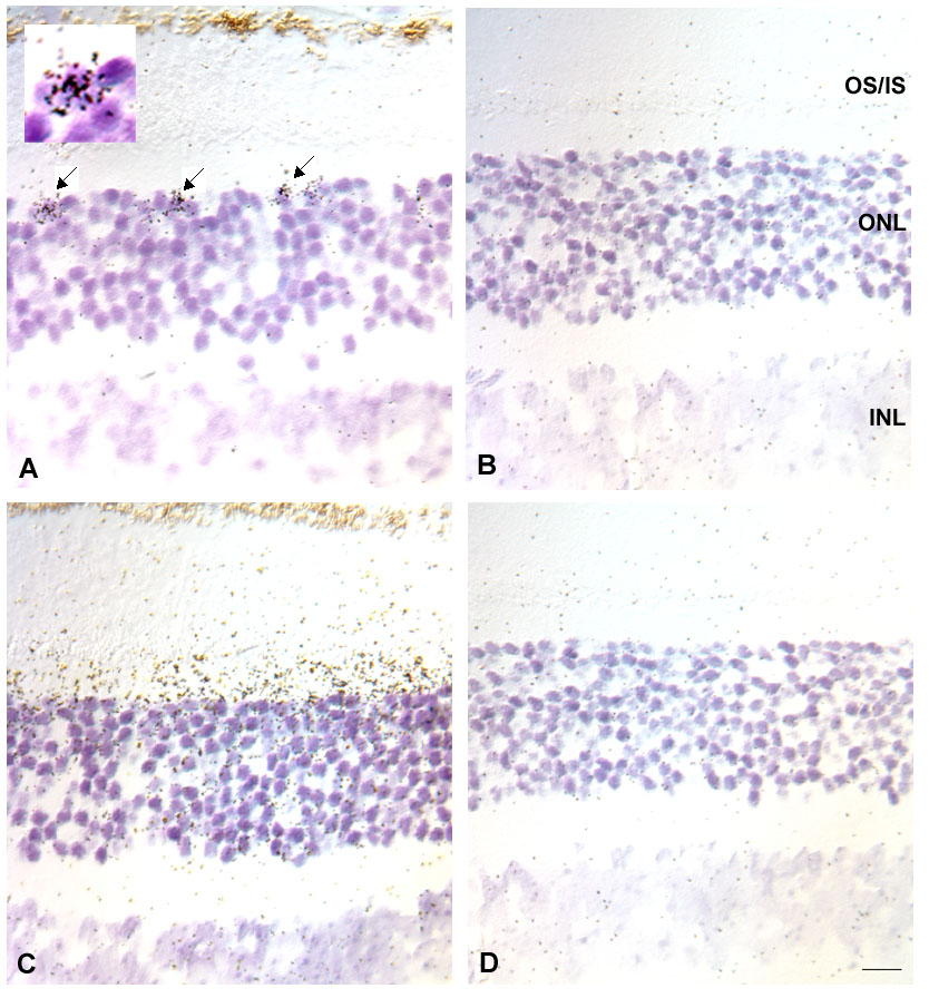
Figure 2. In situ hybridization of mouse retina
In situ hybridization of mouse retina using either the mCAR or mSAG cRNA probes. (A) mCAR antisense cRNA probe. The probe labels a population of cells consistent with the location, appearance, and distribution of cone photoreceptors (arrows). (B) mCAR sense cRNA probe. No specific hybridization signals. (C) mSAG antisense cRNA probe. The probe labels rod inner segment and outer nuclear layers. (D) mSAG sense cRNA probe. Note the lack of hybridization signal over the photoreceptors. OS, outer segments; IS, inner segments; ONL, outer nuclear layer; INL, inner nuclear layer. Bar represents 10 μm.