![]() Figure 5 of
Orem, Mol Vis 2002;
8:455-461.
Figure 5 of
Orem, Mol Vis 2002;
8:455-461.
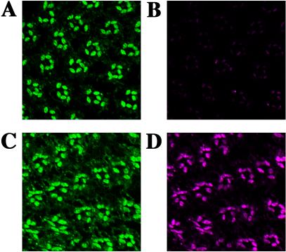
Figure 5. Lowering rhodopsin levels causes epitope masking
Dissected retinas from dark-reared flies were treated with either 15 min of blue light to activate rhodopsin (A and B) or 15 min of orange light to inactivate rhodopsin (C and D), fixed in the dark, and 1 μM sections cut on an ultramicrotome. The sections were stained for rhodopsin using either the Rh1 polyclonal antibody (A and C), or the Rh1 monoclonal antibody (B and D). The brightness level for each image was calibrated using the orange light treated sections, and was not changed for the blue light treated samples. Notice that blue light treatment drastically reduces the amount of rhodopsin accessible to the monoclonal Rh1 antibody (compare B and D).