![]() Figure 2 of
Orem, Mol Vis 2002;
8:455-461.
Figure 2 of
Orem, Mol Vis 2002;
8:455-461.
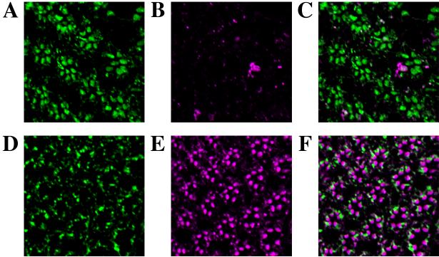
Figure 2. Metarhodopsin immunoreactivity is masked in norpA flies
Flies were treated with 24 h of room light and retinas were dissected. The retinas were then treated with 15 min of either blue light (A-C) to convert rhodopsin to the M form or orange light to convert the rhodopsin to the inactive (R) form (D-F). The treated retinas were then immediately fixed in the dark and 1 μM frozen sections cut on an ultramicrotome. The sections were stained for Arr2 using a polyclonal antipeptide antibody (A and D), and rhodopsin using the monoclonal Rh1 antibody (B and E). Panels C and F show the merged image. Note that after light treatment for 24 h, the rhodopsin monoclonal antibody only recognizes the rhodopsin that has been inactivated by orange light (compare panel B and E).