![]() Figure 1 of
Orem, Mol Vis 2002;
8:455-461.
Figure 1 of
Orem, Mol Vis 2002;
8:455-461.
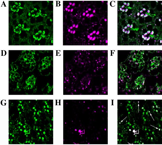
Figure 1. Rhodopsin monoclonal antibody epitope is masked in norpA flies
Frozen (1 μM) sections were cut from wildtype (A, B, C) or norpA (D-I) flies treated with 24 h of room light. Sections were stained for Arr2 using a polyclonal antipeptide antibody (A and D) and rhodopsin using a monoclonal Rh1 antibody (B, E, and H) or a polyclonal Rh1 antibody (G). The white arrows in panel I indicate puncta where both the monoclonal and the polyclonal Rh1 antibodies recognize antigen. Notice in panel G the polyclonal antibody recognizes rhabdomeric rhodopsin, but this rhodopsin is unrecognizable to the monoclonal Rh1 antibody in panel H.