![]() Figure 2 of
Behling, Mol Vis 2002;
8:449-454.
Figure 2 of
Behling, Mol Vis 2002;
8:449-454.
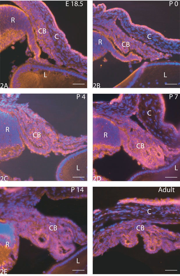
Figure 2. Immunohistochemical localization of PEDF in the ciliary body
Immunohistochemical localization of PEDF in the ciliary body (E18.5-Adult). PEDF was expressed by cells of the ciliary body throughout development (A-F). There were no changes in intensity of PEDF immunofluorescence with developmental age. Nuclei are counterstained with DAPI. Bar represents 50 μm. Abbreviations: CB = ciliary body; C = cornea; R = retina; L = lens.