![]() Figure 1 of
Behling, Mol Vis 2002;
8:449-454.
Figure 1 of
Behling, Mol Vis 2002;
8:449-454.
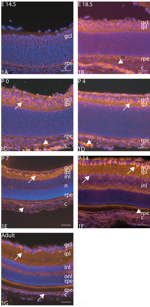
Figure 1. Immunohistochemical examination of PEDF in the neural retina and choroid
Immunohistochemical examination of PEDF in the neural retina and choroid (E14.5-Adult). PEDF was seen in the choroid at all time points shown except E 14.5 (arrowheads). PEDF levels in the ganglion cell layer qualitatively increased with increasing age (arrows; B-F) with highest levels seen at P14 (F). Relative levels decreased slightly at adulthood (G). Nuclei are counterstained with DAPI. Bar represents 50 μm. Abbreviations: gcl = ganglion cell layer; rpe = retinal pigment; c = choroid; inl = inner nuclear layer; ipl = inner plexiform layer; n = neuroblasts; onl = outer nuclear layer.