![]() Figure 1 of
Mukhopadhyay, Mol Vis 2002;
8:442-448.
Figure 1 of
Mukhopadhyay, Mol Vis 2002;
8:442-448.
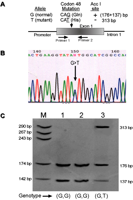
Figure 1. Detection of Gln48His mutation
A: Location of the mutation within MYOC and the nature of the nucleotide change are shown. The allelic difference resulting from the codon 48 polymorphism was determined by Acc I digestion of the PCR product obtained using a pair of primers selected from exon 1 of the gene. B: Representative chromatogram containing sequence from the non coding DNA strand shows the location of the mutation as a double peak in the heterozygous condition (arrow). C: Polyacrylamide gel (6%) analysis of PCR products from three representative samples and their genotypes are shown. Lane M is a pBS/Hae III digested molecular weight marker. The sizes of the molecular weight marker and the Acc I digested DNA fragments are shown in the left and the right sides of the gel, respectively.