![]() Figure 4 of
Kennedy, Mol Vis 2002;
8:422-430.
Figure 4 of
Kennedy, Mol Vis 2002;
8:422-430.
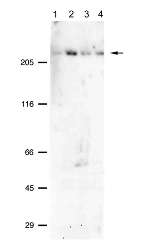
Figure 4. Western blot analysis of P-glycoprotein expression in cultured human RPE using monoclonal antibody mAb JSB-1
All lanes contained 15 μg protein from whole cell lysates of cultured cells. Lanes were loaded as follows: lane 1: cultured human RPE, passage 40; lane 2: cultured human RPE, passage 1; lane 3: D407 cells, passage 57; lane 4: D407 cells, passage 58. Primary antibody was used at final concentration of 0.5 μg/ml. Secondary antibody was anti-mouse IgG conjugated with alkaline phosphatase. Blots were incubated with chemiluminescence substrate (CDP-Star) and the signal digitally captured with a Kodak Image Station 440. A single prominent band is detected at about 225 kD.