![]() Figure 3 of
Kennedy, Mol Vis 2002;
8:422-430.
Figure 3 of
Kennedy, Mol Vis 2002;
8:422-430.
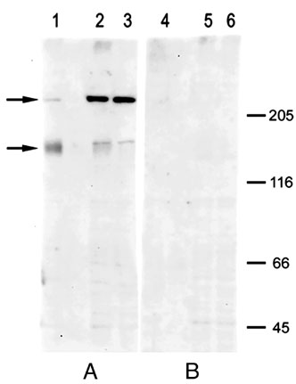
Figure 3. Western blot analysis of P-glycoprotein expression in cultured human RPE using the polyclonal antibody mdr (Ab-1)
Panel A shows reactivity with the antibody (1 μg/ml) plus vehicle, while panel B shows reactivity after preincubation with immunizing peptide (antibody concentration was 1 μg/ml, peptide concentration was 10 μg/ml). Secondary antibody was anti-rabbit IgG conjugated with alkaline phosphatase. Blots were incubated with chemiluminescence substrate (CDP-Star) and the signal digitally captured with a Kodak Image Station 440. Each lane contained 20 μg total protein. Lanes 1 and 4 were loaded with an RPE membrane preparation from cells at passage 11. Remaining lanes contained whole cell lysates from the same human RPE donor culture. Lanes 2 and 5 were from cells at passage 7 while lanes 3 and 6 were from cells at passage 1. Arrows identify bands at 224 and 165 kD.