![]() Figure 1 of
Kennedy, Mol Vis 2002;
8:422-430.
Figure 1 of
Kennedy, Mol Vis 2002;
8:422-430.
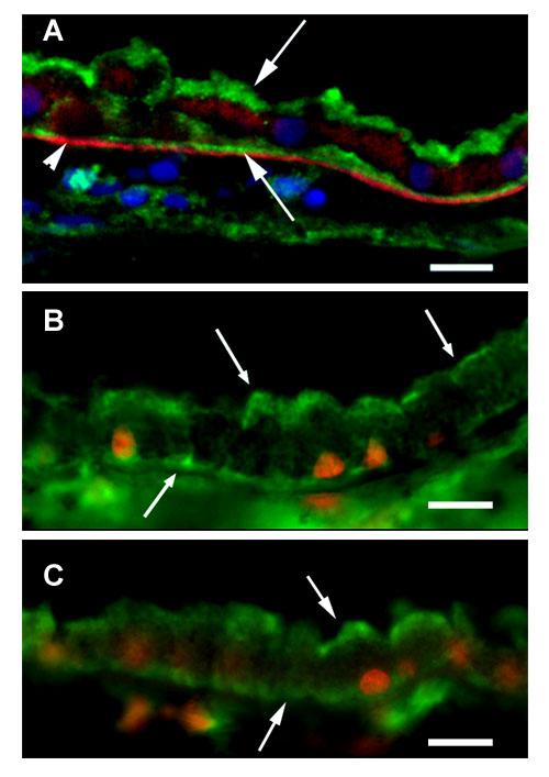
Figure 1. Immunolocalization of P-glycoprotein in RPE/choroid frozen sections
Immunolocalization of P-glycoprotein in frozen sections of native human RPE/choroid using three different antibodies. Tissue orientation in all panels is RPE above choroid. A: Image of RPE/choroid reacted with mAb 4E3. Prominent immunoreactivity (green) is present along the apical surface of the RPE (downward arrow) and also along the basolateral membrane (upward arrow). Arrowhead indicates Bruch's membrane, which exhibits modest autofluorescence (red). B: RPE/choroid section reacted with mAb UIC2. C: Section reacted with mAb JSB-1. As in A, prominent immunoreactivity is distributed along both the apical and basolateral RPE cell membranes. Scale bars in all panels represent 20 μm.