![]() Figure 8 of
Vittitow, Mol Vis 2002;
8:32-44.
Figure 8 of
Vittitow, Mol Vis 2002;
8:32-44.
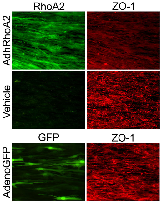
Figure 8. AdhRhoA2 induced changes in ZO-1 distribution in SC cells
Monolayers of 4-day confluent SC cells were infected with 500 pfu/cell AdhRhoA2. After 48 h, cells were fixed, permeablized and double-stained with specific antibodies against RhoA and ZO-1 using Cy2 (RhoA) and Cy3 (ZO-1) conjugated secondary antibodies. ZO-1 staining is present in the vehicle-treated cells at the cell-cell junctions and appear as a segmented border. Cells infected with AdhRhoA2 are devoid of this apportioned ZO-1 staining at the intercellular junctions. Original magnification noted at the right of the microphotographs.