![]() Figure 7 of
Vittitow, Mol Vis 2002;
8:32-44.
Figure 7 of
Vittitow, Mol Vis 2002;
8:32-44.
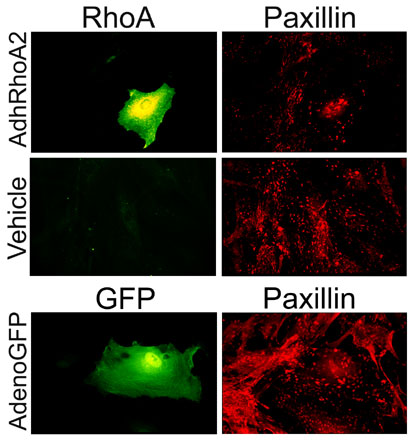
Figure 7. Immunofluorescent localization of paxillin in HTM cells infected with AdhRhoA2
Sub-confluent cultures of HTM cells were infected with 100 pfu/cell of AdhRhoA2 and AdenoGFP. At 48 h post-infection, cells were fixed, permeablized and processed for immunofluorescence. Cells treated with AdhRhoA2 and vehicle were double-stained with specific antibodies for RhoA and paxillin using FITC (RhoA) and TRITC (paxillin) conjugated secondary antibodies. Cells infected with AdenoGFP were stained for paxillin using a TRITC-conjugated secondary antibody. Panels were visualized with the fluorescein (left, RhoA and GFP) and rhodamine (right, paxillin) channels. Paxillin staining is reduced in AdhRhoA2 infected cells while it is essentially unchanged in cells infected with AdenoGFP. Original magnification 100x.