![]() Figure 3 of
Vittitow, Mol Vis 2002;
8:32-44.
Figure 3 of
Vittitow, Mol Vis 2002;
8:32-44.
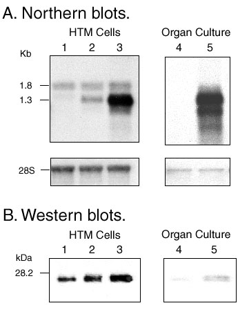
Figure 3. Adenoviral gene transfer of dominant-negative RhoA
A: Northern blots of total RNA extracted from cultures infected with AdhRhoA2 and hybridized to a full-coding RhoA cDNA probe. Left: HTM cells infected at 10 and 100 pfu/cell and harvested for RNA 48 h post-infection. Right: TM tissue obtained from the perfused anterior segment of a pair of eyes where one eye was injected with 107 pfu of AdhRhoA2 and the contralateral eye was injected with vehicle. Tissue was dissected 48 h post-injection. Lane 1: Vehicle. Lane 2: 10 pfu/cell. Lane 3: 100 pfu/cell. Lane 4: Vehicle. Lane 5: AdhRoA2.
B: Western Blots of proteins extracted from cultures infected with AdhRhoA2 probed with a rabbit anti-human RhoA antibody and visualized by chemiluminescence. Left: equivalent volumes of extracts from HTM cells infected at 10 and 100 pfu/cell and harvested 48 h post-infection. Right: Protein extract from a whole TM tissue obtained from the perfused anterior segment of a pair of eyes where one eye was injected with 107 pfu of AdhRhoA2 and the contralateral eye was injected with vehicle. Tissue was dissected 48 h post-injection. A dose-dependent increase in RhoA mRNA and protein was observed in the infected cells as well as the intact human trabecular meshwork after a single dose injected though the cornea of the perfused anterior segment. Lanes as described in A.