![]() Figure 7 of
Tasheva, Mol Vis 2002;
8:407-415.
Figure 7 of
Tasheva, Mol Vis 2002;
8:407-415.
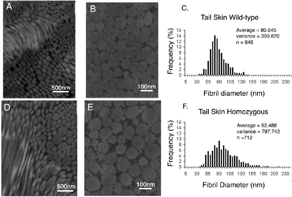
Figure 7. TEM of collagen fibrils from mouse tail skin
Transmission electron micrographs containing cross sections of collagen fibrils from tail skin of wild type mouse (A and B) and mimecan-null mouse (D and E). Morphometric analysis of tail skin fibrils (C and F). Notice that the range of distribution is larger in mimecan-null mice (F).