![]() Figure 6 of
Tasheva, Mol Vis 2002;
8:407-415.
Figure 6 of
Tasheva, Mol Vis 2002;
8:407-415.
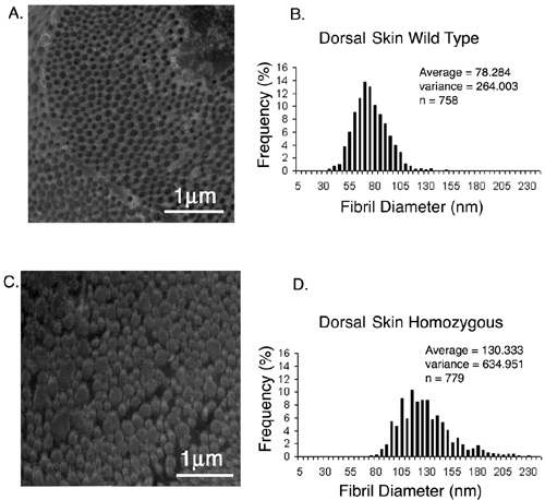
Figure 6. TEM of collagen fibrils from mouse dorsal skin
Transmission electron micrographs containing cross sections of collagen fibrils from dorsal skin of wild type mouse (A) and mimecan-null mouse (C). Morphometric analysis of skin collagen fibrils (B and D). Notice the significantly larger diameter of collagen fibrils in mimecan-null mice (D).