![]() Figure 5 of
Tasheva, Mol Vis 2002;
8:407-415.
Figure 5 of
Tasheva, Mol Vis 2002;
8:407-415.
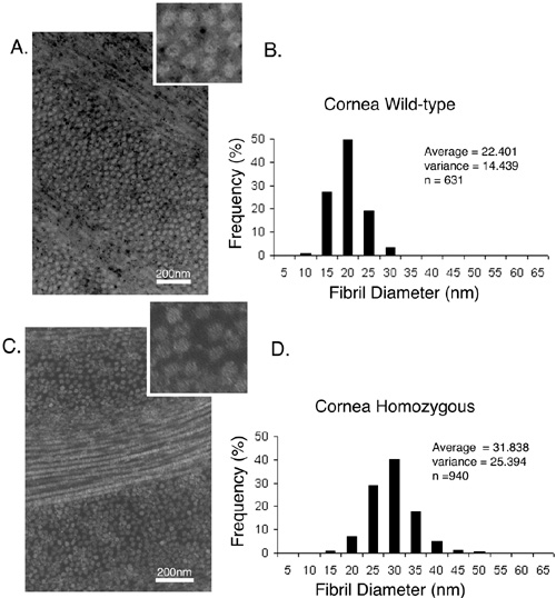
Figure 5. TEM of collagen fibrils from mouse corneal stroma
Transmission electron micrographs containing cross-sections of collagen fibrils from corneal stroma from wild-type (A) and mutant (C) mice. Morphometric analysis of corneal collagen fibrils in wild-type (B) and in mutant (D) mice. Fibril diameter was measured as described in methods and presented in a histogram. The average fibril diameter, the variance and the total number (n) of fibrils measured are indicated.