![]() Figure 4 of
Tasheva, Mol Vis 2002;
8:407-415.
Figure 4 of
Tasheva, Mol Vis 2002;
8:407-415.
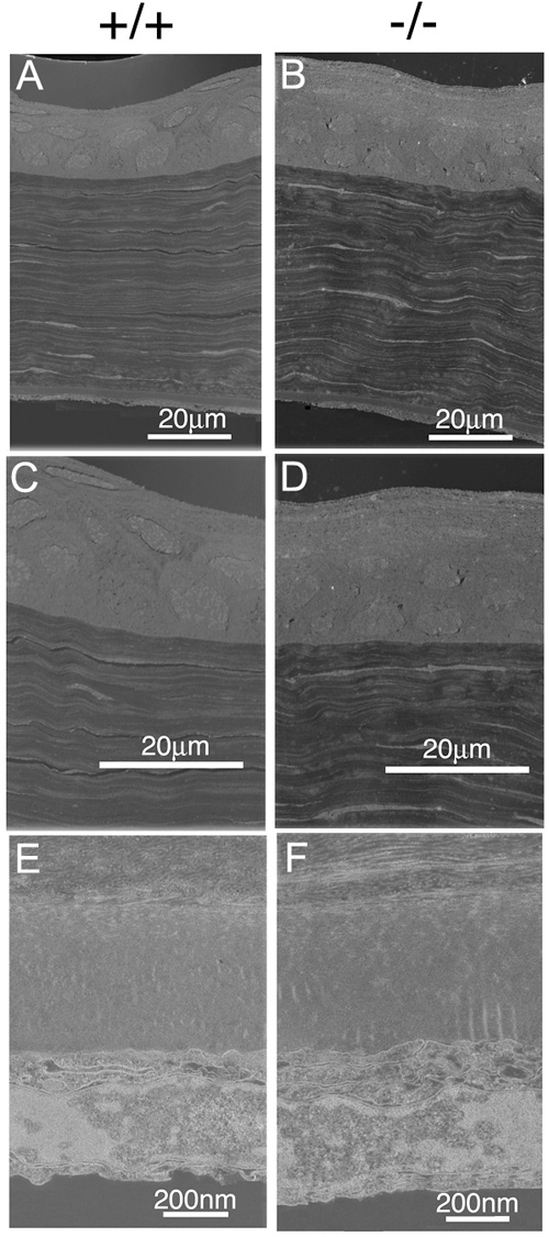
Figure 4. TEM of corneas
Transmission electron micrographs of corneas from wild type (+/+) and mimecan-null mice (-/-). A and B: Longitudinal sections of the cornea; C and D: Higher magnification of the epithelial sublayer of the cornea; E and F: Descemet's and endothelial layers of the cornea. No significant differences in the cornea and corneal layers can be noted.