![]() Figure 7 of
Chen, Mol Vis 2002;
8:372-388.
Figure 7 of
Chen, Mol Vis 2002;
8:372-388.
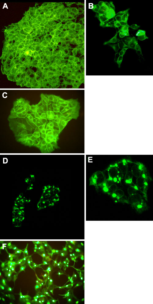
Figure 7. Expression of MP19green and MP19To3 green in 293 cells
T-RexTM-293 (293) cells were transfected with either the MP19-pEGFP-pcDNA4/TO (MP19greenTO) or MP19To3-EGFP-pcDNA4/TO (MP19To3greenTO) vectors and stable clones were picked following Zeocin selection. The stable cell line containing MP19greenTO or MP19To3greenTO was seeded onto coverslips and induced with tetracycline when the cells reached the correct cell density. Fluorescent cells were then observed with both confocal and Epi-fl microscopy. A: Confocal microscopy analysis of MP19green expression in 293 cells (EGFP filter set). The image was obtained using the inverted microscope with a 40x water objective. B: Epi-fl microscopy analysis of MP19green expression in 293 cells (EGFP filter set). The image was obtained using a Nikon Optiphot-2 upright microscope with the episcopic-fluorescence attachment EFD-3. Cells were observed using a 40x oil objective. C: Conditions were identical with those in B, view of a different area of the coverslip. D: Confocal microscopy analysis of MP19To3green expression in 293 cells (EGFP filter set). The image was obtained using the inverted microscope with a 40x water objective. E: Conditions were identical with those in D, view of a different area of the coverslip. F: Epi-fl microscopy analysis of MP19To3green expression in 293 cells (EGFP filter set). The image was obtained using a Nikon Optiphot-2 upright microscope with the episcopic-fluorescence attachment EFD-3. Cells were observed using a 40x oil objective.