![]() Figure 3 of
Chen, Mol Vis 2002;
8:372-388.
Figure 3 of
Chen, Mol Vis 2002;
8:372-388.
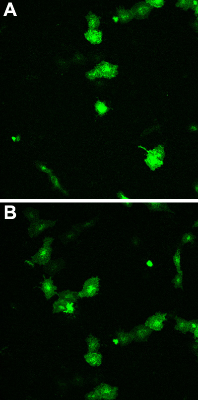
Figure 3. MP19-EGFP expression in CLE
CLE cells were transfected with vector MP19-pEGFP-N1. About 24 h later, fluorescent protein was observed using the confocal microscope. A and B are views of different cultures taken on separate days. The fluorescent MP19/EGFP protein appears to migrate to the cell membrane and collect in a punctate fashion on the membrane. The image was obtained using the inverted microscope with a 40x water objective.