![]() Figure 18 of
Chen, Mol Vis 2002;
8:372-388.
Figure 18 of
Chen, Mol Vis 2002;
8:372-388.
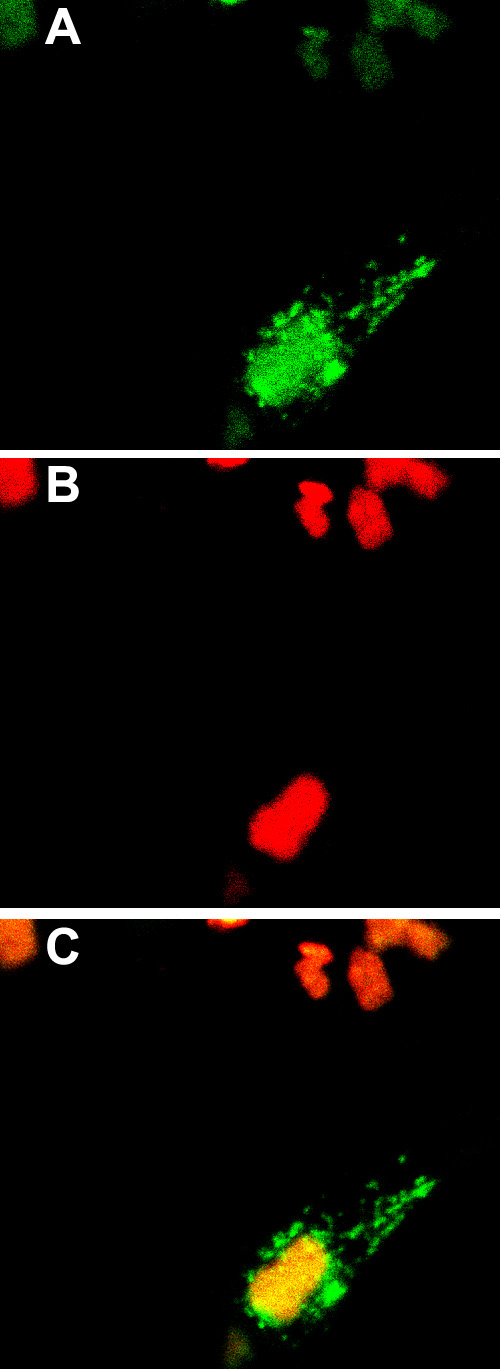
Figure 18. MP19To3red and Golgi-green protein expression in 293 cells
MP19To3redTO cells were cotransfected with the Golgi-pEGFP-N1 plasmid. 48 h following transfection with Golgi-pEGFP-N1, the cells were induced with tetracycline. The cells were observed with confocal microscopy (Both EGFP and DsRed filter sets). A: Image of cells using the EGFP filter set (Golgi-green). B: Image of cells using the DsRed filter set (MP19To3red). C: Superimposed image from A and B. The image was obtained using the inverted microscope with a 40x water objective. The images were further magnified using Adobe Photoshop 7.0.