![]() Figure 15 of
Chen, Mol Vis 2002;
8:372-388.
Figure 15 of
Chen, Mol Vis 2002;
8:372-388.
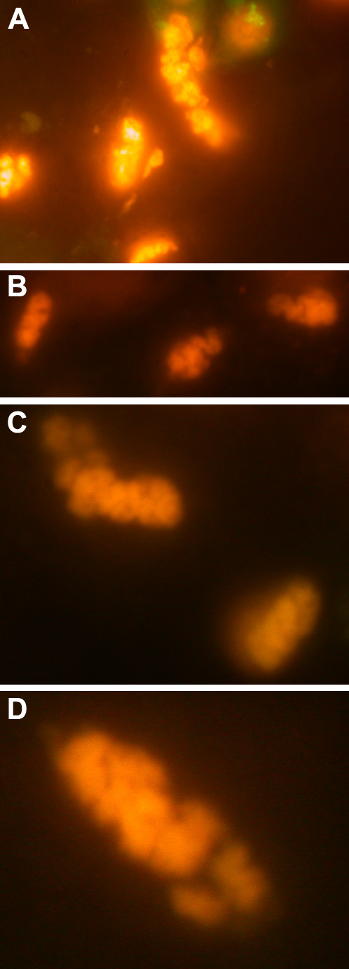
Figure 15. Observation of MP19To3red protein inclusion bodies in 293 cells
MP19To3redTO cells were induced with tetracycline when the cells reached the correct cell density. About 7-10 days following transfection, the cells were observed using Epi-fl microscopy (DsRed filter set). A, B, C, and D are magnified views of different MP19To3red protein inclusion bodies in various 293 cells. It is obvious that the nature of the inclusion bodies are of a tubular, twisting shape. The images were obtained using a Nikon Optiphot-2 upright microscope with the episcopic-fluorescence attachment EFD-3. Cells were observed using a 60x oil objective. The cells observed in all of the images were photographed using a Nikon Coolpix 995 digital camera.