![]() Figure 12 of
Chen, Mol Vis 2002;
8:372-388.
Figure 12 of
Chen, Mol Vis 2002;
8:372-388.
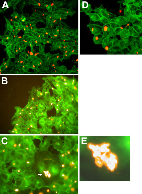
Figure 12. MP19green and MP19To3red protein coexpression in 293 cells
The MP19greenTO cell line was seeded onto coverslips and cotransfected with MP19To3-pDsRed2-N1, which expressed MP19To3 fused to the red fluorescent protein, DsRed. After 48 h following transfection with MP19To3pDsRed2-N1, the cells were induced with tetracycline and observed for fluorescence 24 h later. A: Analysis of MP19green and MP19To3red coexpression in 293 cells, using confocal microscopy with both the EGFP and DsRed filter sets. The image was obtained using the inverted microscope with a 40x water objective. B: Analysis of MP19green and MP19To3red coexpression in 293 cells, using Epi-fl microscopy (EGFP and DsRed filter sets). The image was obtained using a Nikon Optiphot-2 upright microscope with the episcopic-fluorescence attachment EFD-3. Cells were observed using a 40x oil objective. C: Conditions were identical with those in B, view of a different area of the coverslip and the image was magnified 2-fold. Note the "giant" cell in the center of the image, with a very large inclusion of MP19To3red protein (arrow). D: Conditions were identical with those in A, view of a different area of the coverslip and the image was magnified 3-fold. E: Magnified view of the very large inclusion body containing MP19To3red protein observed in C, arrow.