![]() Figure 5 of
Li, Mol Vis 2002;
8:341-350.
Figure 5 of
Li, Mol Vis 2002;
8:341-350.
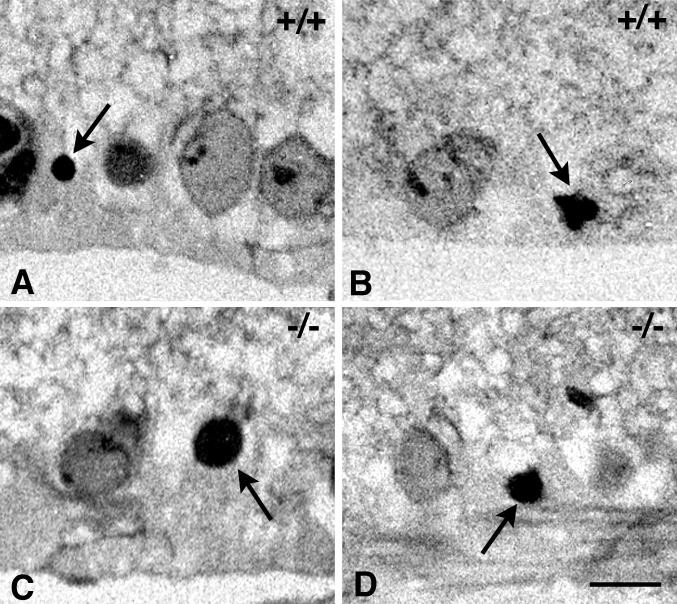
Figure 5. Histologic evaluation of the ganglion cell layer in NMDA treated p53 mutant mice
Cell death in p53 wild-type and p53-null mice is associated with the formation of pyknotic nuclei. Micrographs of toluidine blue-stained retinal sections taken 2 days after injection of 160 nmol of NMDA. A, B: Close-up of the ganglion cell layer of wild-type mice showing both normal appearing and pyknotic nuclei (arrows). C, D: Close-up of the ganglion cell layer of p53-null mice showing similar pyknotic nuclei. Based on morphological criteria, these results suggest that an apoptotic-like mechanism is active in both groups of mice. Size bar represents 7 μm.