![]() Figure 1 of
Li, Mol Vis 2002;
8:341-350.
Figure 1 of
Li, Mol Vis 2002;
8:341-350.
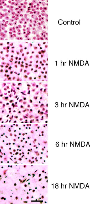
Figure 1. Effect of NMDA on nuclear morphology in the ganglion cell layer
Changes in nuclear morphology are rapidly observed in cells of the ganglion cell layer after intravitreal injection of NMDA. Shown are Nissl-stained wholemounts of mouse retinas harvested at 1, 3, 6, and 18 h after intravitreal injection of 40 nmol of NMDA. At 1 h, several cells in the ganglion cell layer show early signs of changes in nuclear morphology, suggestive of early stages of chromatin condensation. By 3 h, nearly all the presumed RGCs have this appearance or have become pyknotic. All these cells are pyknotic by 6 h. After 18 h, few cells remain. For the most part, these remaining cells and their nuclei have become fragmented and appear to be in the final stages of death. Size bar represents 35 μm.