![]() Figure 4 of
Smith, Mol Vis 2002;
8:26-31.
Figure 4 of
Smith, Mol Vis 2002;
8:26-31.
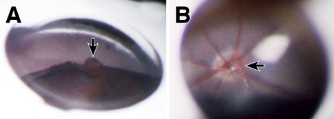
Figure 4. Additional uses of goniolens
A: DBA/2J mouse, 12 months of age. In a slightly oblique view through the center of the goniolens (corneal contact lens), an elevation of the iris surface due to a focal collection of pigmented cells (arrow) is present at the pupillary margin. B: The optic nerve, retinal vessels, and posterior pole retina of a normal two month old C57BL/6J mouse as seen through the center of the goniolens. The nerve is indicated by the arrow. The retinal vessels pass over the edge with a slight curvature that indicates the shallow physiological cup.