![]() Figure 3 of
Smith, Mol Vis 2002;
8:26-31.
Figure 3 of
Smith, Mol Vis 2002;
8:26-31.
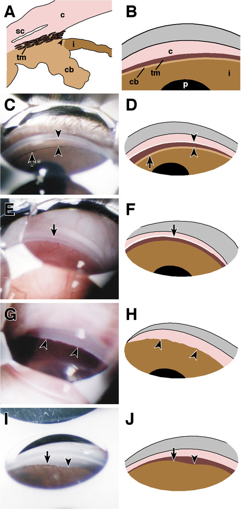
Figure 3. Goniophotographs of normal and abnormal angles
A: Schematic cross-section of a mouse eye showing the angle region. Indicated structures are the cornea (c), Schlemm's canal (sc), trabecular meshwork (tm), iris (i), and ciliary body (cb). The region immediately next to the iris root that is often referred to as the ciliary band is marked by the arrowhead. The same colors are used to designate these structures throughout this figure and are labeled only where needed for clarity. In the schematic gonioscopic views (B, D, F, H, J) Schlemm's canal is represented by a white band and the most peripheral cornea in pink. The upper gray area represents the out of focus image of more central corneal tissue and other surface structures that always appear at this location when performing gonioscopy on mice or humans. B: Schematic gonioscopic view of the normal mouse angle. A portion of the anterior surface of the ciliary body (cb, often referred to as the ciliary band) is sometimes apparent between the trabecular meshwork and iris, but is not always evident. Schlemm's canal is not represented in this schematic. The visibility of Schlemm's canal depends on contrast provided by pigmentation or by the presence of blood in the canal. Occasionally, a reflux of blood into Schlemm's canal also occurs during human gonioscopy [37]. "p" indicates the pupil. C: Normal adult C57BL/6J angle. The lightly pigmented trabecular meshwork is located between the arrowheads. The tip of the upper arrowhead touches Schlemm's canal, which appears white. Due to the presence of blood in a similar structure at the same location during some examinations, we interpret this structure to be Schlemm's canal and not Schwalbe's line. A small portion of the face of the ciliary body is evident (arrow). D: Schematic gonioscopic view corresponding to panel C. E: Adult albino C57BL/6J mouse (C57BL/6JTyrc-2J/c-2J). Though not evident in the photograph blood was visible in Schlemm's canal. Schlemm's canal is indicated by the arrow. The TM is evident, but appears semitransparent due to lack of pigmentation. F: Schematic gonioscopic view corresponding to panel E. Schlemm's canal is indicated by the white line. The ciliary body is not seen because of the lack of pigment. The iris and trabecular meshwork are depicted with the same colors as those used for pigmented mice to keep the diagram analogous with the others. Panels G-J demonstrate anterior synechiae in 11 month old DBA/2J mice. The synechiae are of different severity as is reminiscent of the disease in this strain [10]. G: Pigmented broad anterior synechiae (arrowheads) conceal the trabecular meshwork and have a scalloped appearance. H: Schematic gonioscopic view corresponding to panel G. I: Anterior synechiae have progressed more centrally and obscured more of the trabecular meshwork on the left side of the view (arrow) than on the right (arrowhead). J: Schematic gonioscopic view corresponding to panel I.