![]() Figure 2 of
Smith, Mol Vis 2002;
8:26-31.
Figure 2 of
Smith, Mol Vis 2002;
8:26-31.
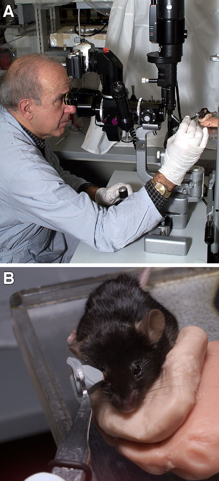
Figure 2. Use of goniolens
A: Gonioscopy on an anesthetized mouse. The operator's hand rests for stability on the modified slit lamp platform. B: Close-up view shows the goniolens in contact with the cornea. The mouse is supported and positioned on a moldable platform to avoid movement. Stainless steel holders [18] keep the mouse in position (not visible).