![]() Figure 7 of
Fei, Mol Vis 2002;
8:306-314.
Figure 7 of
Fei, Mol Vis 2002;
8:306-314.
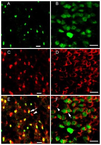
Figure 7. PNA labeling of the cones in the rd1 retinas
Double-labeling of the fluorescent cells expressing the GFP transgene in the developing and mature rd1 retinas by cone cell-specific PNA marker. These confocal images were taken from PNA-stained retinal wholemounts. A, C, and E were from a P7 rd1 mouse with microscope focusing on the base of the cone outer segments. B, D, and F were from an adult rd1 mouse with focusing on the cone cell bodies. A and B were FITC images (pseudocolored green) of the fluorescent cone photoreceptor cells. C and D were rhodamine images (pseudocolored red). E and F were the merged images, showing that all green fluorescent cells in the developing and adult rd1 retinas were stained by PNA (arrows), although not all cones expressed the GFP (arrowheads). This staining confirmed that the green fluorescent cells in the developing and adult rd1 mouse retinas were the cones. Scale bar: 10 μm.