![]() Figure 6 of
Fei, Mol Vis 2002;
8:306-314.
Figure 6 of
Fei, Mol Vis 2002;
8:306-314.
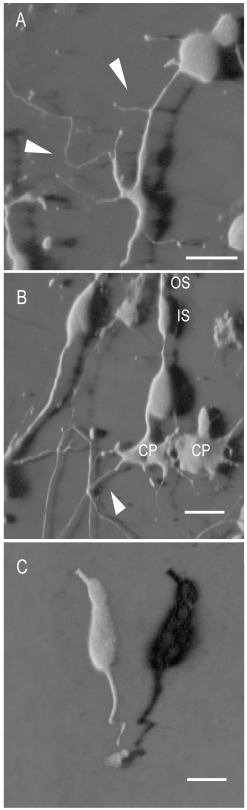
Figure 6. Robust abnormal neurite sprouting from the cone axons and pedicles in the P10 rd1 retinas
Cone neurite sprouting and branching in the rd1 retina became more evident by P10 (A and B, arrowheads). Some cone pedicles projected long, branching, filopodia-like processes, while in the control, the cones did not show neurite sprouting (D). The cones appeared to maintain a grossly normal cone-cone contact through their pedicles (B, CP) and the sprouting seemed to occur from the sites of a cone pedicle that did not contact the neighboring pedicle. The neurite sprouting occurred even when the cone still appeared to preserve a grossly normal morphology of the outer and inner segment (B, OS and IS). All images were 3D shadow projections of confocal microscopic images from P10 mouse retinal wholemounts. Scale bar 10 μm.