![]() Figure 5 of
Fei, Mol Vis 2002;
8:306-314.
Figure 5 of
Fei, Mol Vis 2002;
8:306-314.
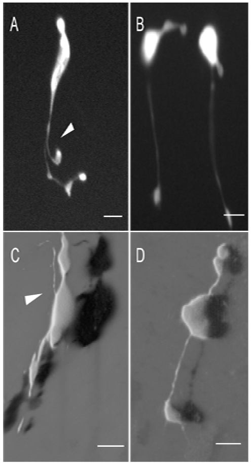
Figure 5. Abnormal cone neurite sprouting in the rd1 mice began at P8
Apparent cone neurite outgrowth arose from the cone axons of the P8 rd1 mouse retinas (A and C, arrowheads), which was not noticed in the normal cones of the control (B and D). A and B were epifluorescence microscopic images of the cones in live retinal wholemounts. C and D were 3D shadow projections of confocal images of the fluorescent cones in fixed retinal wholemounts. Scale bar: 10 μm.