![]() Figure 4 of
Fei, Mol Vis 2002;
8:306-314.
Figure 4 of
Fei, Mol Vis 2002;
8:306-314.
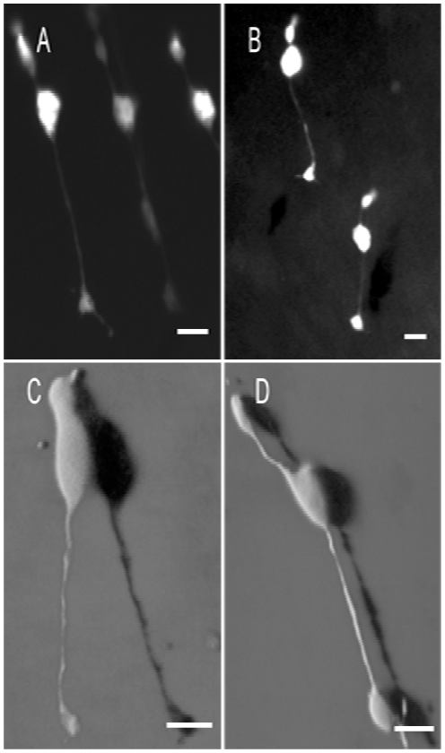
Figure 4. Cones appeared to be normal by P7
Cone cells appeared to be morphologically normal by P7 in rd1 mice. The morphology of cones in the rd1 mouse retinas at P7 (A and C) appeared to be comparable to that of the control retinas (B and D). The cone outer segments were still developing. Although some cone pedicles exhibited prominent processes in the rd1 retinas, these processes were also present in the controls, and no abnormal neurite sprouting was observed. A and B were epifluorescence microscopic images of the cones in live retinal wholemounts. C and D were 3D shadow projections of confocal images of the fluorescent cones in fixed retinal wholemounts. Scale bar: 10 μm.