![]() Figure 3 of
Fei, Mol Vis 2002;
8:306-314.
Figure 3 of
Fei, Mol Vis 2002;
8:306-314.
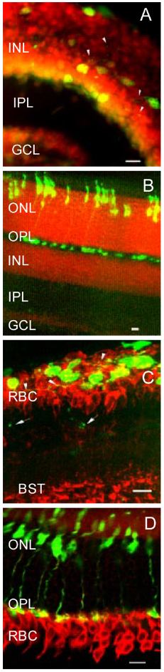
Figure 3. Migration of degenerating cones and projection of cone neurites
Migration of the degenerating cones into the inner nuclear layer (INL) and projection of cone neurites to the inner plexiform layer (IPL) in the rd1 retinas. Counterstaining of the retinal sections with propidium iodide (A and B) showed the migration of the degenerating cones from the markedly thinning ONL into the INL and the projection of cone neurites toward different directions (A, arrowheads). In the control (B), the cone somata were restricted within the ONL and the cone pedicles within the outer plexiform layer (OPL). Immunostaining of rod bipolar cells (RBC) with anti-PKC-alpha antibody (C) confirmed that the degenerating cones migrated into the INL and that some cone neurites extended horizontally and appeared to contact the RBCs (C, arrowheads), while others extended vitreally past the RBC somata (C, arrows) and reached the IPL. In the control retina immunostained with the same antibody (D), cone somata were located within the ONL and the cone pedicles as well as the RBC dendrites lay in the OPL. GCL: ganglion cell layer. BST: synaptic terminals of rod bipolar cells. Scale bar: 10 μm.