![]() Figure 2 of
Fei, Mol Vis 2002;
8:306-314.
Figure 2 of
Fei, Mol Vis 2002;
8:306-314.
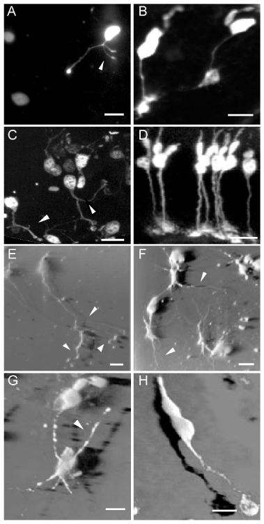
Figure 2. Degenerating rd1 cones undergo neurite sprouting
Degenerating cones in the adult rd1 mouse retinas underwent a remarkable process of neurite sprouting. These cones sprouted neurites (arrowheads) that could be readily identifiable in both live unfixed (A) and fixed (C, E, F, and G; arrowheads) adult rd1 retinas. The abnormal cone neurite had beaded varicosity, and often terminated as a club-shaped structure. The neurites were budding from the degenerating cones and outgrowing in different directions. These abnormal cone processes were evident in 3D view of confocal images of the cones in rd1 mouse retinas (E, F, and G). Some cone pedicles projected long, filopodia-like processes (F and G, arrowheads). The cone pedicle in the control had much shorter processes. In the rd1 retinas, some cones lost their outer and inner segments (A) and became disorganized (C), while others lost all their processes and left only dead cell bodies that showed diminished fluorescence (A). All these abnormalities of cones were not observed in the corresponding controls (B, D, and H). The normal morphology of a typical cone cell, including the outer and inner segments, cell soma, axon and a large, flattened synaptic pedicle, can be readily appreciated in D and H. A to D were epifluorescence microscopic images from mice aged 2 months. A and B were from live retinal wholemounts, and C and D were from fixed retinal sections. E, F, G, and H were 3D shadow projections of confocal images of the fluorescent cones from fixed retinal wholemounts of mice aged 1 month. Scale bar: 10 μm.