![]() Figure 3 of
Venkatesh, Mol Vis 2002;
8:294-297.
Figure 3 of
Venkatesh, Mol Vis 2002;
8:294-297.
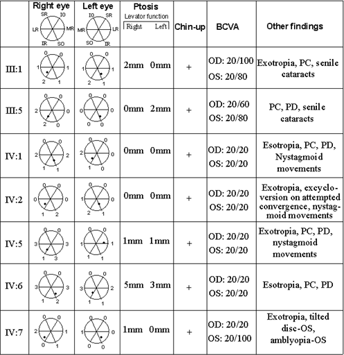
Figure 3. The phenotype and diagrammatic representation of extraocular muscle range of movement
The clinical phenotype and diagrammatic representation of right and left eye extraocular muscle range of movement. Black dot indicates the pupillary position with the head held straight. Movements are measured from each eye's primary resting position. IO, inferior oblique; IR, inferior rectus; SR, superior rectus; LR, lateral rectus; MR, medial rectus; SO, superior oblique; 3, full range of movement; 2, moderate restriction; 1, marked restriction; o, no movement.
