![]() Figure 7 of
Stenkamp, Mol Vis 2002;
8:280-293.
Figure 7 of
Stenkamp, Mol Vis 2002;
8:280-293.
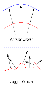
Figure 7. Schematic of vectorial growth in the normal and regenerating retina
Top: In the normal retina growth occurs in an annular fashion, with new cells born at the circumferential germinal zone (red line) and appositionally added to the extant retina, forming a new retinal margin (blue line) that is in spatial register with the old margin. The direction of growth (black arrows), which is locally perpendicular to the tangent of the retinal edge, is spatially aligned with the pre-existing patterns of retinal cells (black dotted lines). Bottom: In this model the edge of a lesion site is quite "jagged" compared to the retinal margin. The vectors of regenerative growth (black arrows), which extend into the lesion site, may be spatially and temporally uncorrelated, and may also be uncorrelated with the pre-existing cellular patterns in extant retina (black dotted lines). These spatiotemporal abnormalities in growth are hypothesized to contribute to the atypical cellular patterns of regenerated retina.