![]() Figure 6 of
Stenkamp, Mol Vis 2002;
8:280-293.
Figure 6 of
Stenkamp, Mol Vis 2002;
8:280-293.
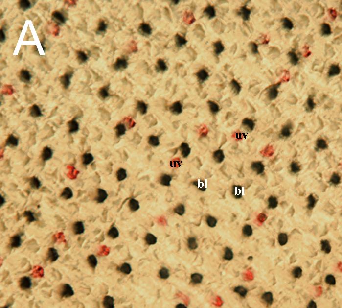
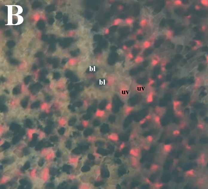
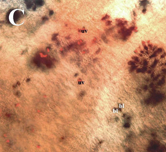
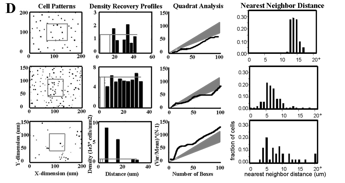
Figure 6. Unusual cone patterns and ratios in goldfish retina following a ouabain-induced lesion
A: Cone mosaic of native goldfish retina. Retina was hybridized with cRNA probes corresponding to blue cone opsin (bl, dark color product) and UV cone opsin (uv, red color product). B: Cone mosaic of peripheral goldfish retina, which had been generated after destruction of the retina by ouabain. C: Central region of the same retina, which had regenerated from stem cells that survived the toxin. D: Analysis of blue cone pattern in native (top row), peripheral (middle row) and regenerated (bottom row) goldfish retina following intraocular injection of ouabain. First column contains cartoons of the 2-dimensional pattern of cones, second column contains DRP analytical results, third column contains results from quadrat analysis, and fourth column contains NND histograms. Figure adapted from Stenkamp et al. [65].
Modified and reproduced with permission from: Stenkamp DL, Powers MK, Carney LH, Cameron DA. Evidence for two distinct mechanisms of neurogenesis and cellular pattern formation in regenerated goldfish retinas. Journal of Comparative Neurology 2001; 431:363-81. Copyright 2001, John Wiley and Sons, Inc.