![]() Figure 5 of
Stenkamp, Mol Vis 2002;
8:280-293.
Figure 5 of
Stenkamp, Mol Vis 2002;
8:280-293.
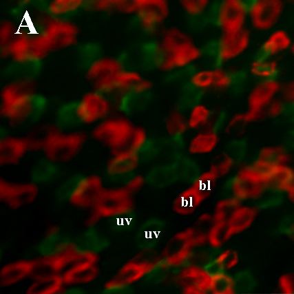
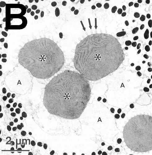
Figure 5. Unusual cone patterns and phenotypes in regenerated retina
A: Cone mosaic of regenerated zebrafish retina, following a surgical lesion. Retina was hybridized with cRNA probes corresponding to blue cone opsin (bl, red fluorescence) and UV cone opsin (uv, green fluorescence). B: A triple cone, an anomalous cone morphology, in the regenerated retina of a sunfish [83]. Asterisks, the outer segments of each triple cone member; A, accessory outer segments; arrows, calical processes. The black profiles are melanin granules within projections that arise from the pigmented epithelium. Anomalous, multiple-ordered cone morphologies of this type are common in the regenerated retina of teleosts.
Panel B is reproduced with permission from: Cameron DA, Easter SS Jr. Cone photoreceptor regeneration in adult fish retina: phenotypic determination and mosaic pattern formation. J Neurosci 1995; 15:2255-71. Copyright 1995, Society for Neuroscience.