![]() Figure 1 of
Stenkamp, Mol Vis 2002;
8:280-293.
Figure 1 of
Stenkamp, Mol Vis 2002;
8:280-293.
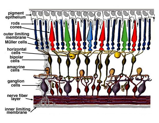
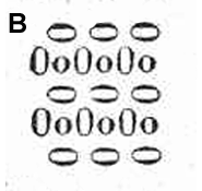
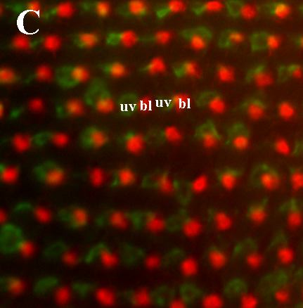
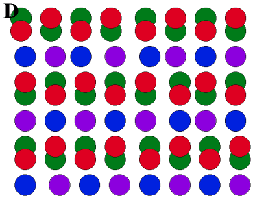
Figure 1. Cellular organization of the vertebrate retina
A: Laminar arrangement of vertebrate retinal cells; schematic obtained from Webvision, The Organization of the Vertebrate Retina, with permission. B: Schematic of the cone photoreceptor mosaic of Perca fluviatilis (European perch) from p. 329 of Hannover [5]. The cylindrical profiles represent double cones ("Zwillingzapfen"), and the circular profiles represent single cones. We believe this to be the first published description and illustration of a teleost cone mosaic. C,D: Cone mosaic of zebrafish. C: A fragment of whole retina hybridized with cRNA probes corresponding to the opsin messages for S ("blue") cones (bl, red fluorescence) and UV cones (uv, green fluorescence). D: A schematic of the zebrafish cone mosaic, illustrating the spatial relationship of the different spectral cone types. Purple, UV cones; Blue, "blue" cones; Green, "green" cones; Red, "red" cones. The juxtaposed "red" and "green" cones constitute the double cone morphology.
A