![]() Figure 1 of
Mukhopadhyay, Mol Vis 2002;
8:271-279.
Figure 1 of
Mukhopadhyay, Mol Vis 2002;
8:271-279.
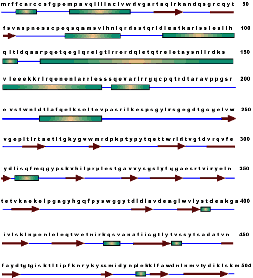
Figure 1. Predicted secondary structure of human myocilin
Open rectangle, solid arrows, and straight lines represent α-helix, β-sheet and turn, and coiled-coils, respectively. The N-terminal half is primarily α-helix; the C-terminal half contains mostly β-sheet and turn, and coiled-coils.