![]() Figure 1 of
Lee, Mol Vis 2002;
8:235-240.
Figure 1 of
Lee, Mol Vis 2002;
8:235-240.
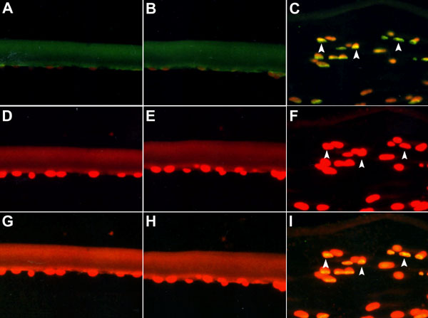
Figure 1. Detection of cell death by TUNEL staining in human anterior polar cataracts
The treated sections were viewed under an epifluorescence microscope. In non-cataractous clear lens, cells were stained with propidium iodide (D) but not with fluorescein (A). There was no co-localization of fluorescein and propidium iodide (G). In nuclear cataracts, cells were stained with propidium iodide (E) but not with fluorescein (B). There was no co-localization of fluorescein and propidium iodide (H). A subpopulation of apoptotic cells, scattered throughout the tissue section from anterior polar cataracts, were intensely stained (bright green to yellow, C) with fluorescein. Cells were counterstained with propidium iodide which stains both apoptotic and nonapoptotic cells (red, F). The co-localization of fluorescein and propidium iodide resulted in a bright yellow mixed color by double filter analysis (I). Arrows point to areas of apoptosis, where fluorescein and propidium iodide were co-localized. The data presented was from one of six independent assays that produced similar results. The original magnification was 200 x.