![]() Figure 8 of
Lee, Mol Vis 2002;
8:17-25.
Figure 8 of
Lee, Mol Vis 2002;
8:17-25.
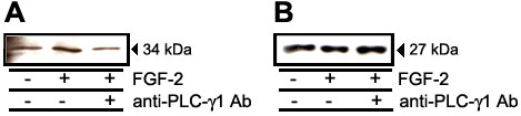
Figure 8. Effect of FGF-2 and anti-PLC-g1 antibody on Cdk4 and p27Kip1
When CEC reached approximately 80% confluency, they were replaced in serum-free medium for 24 h. Cells were then treated with FGF-2 (10 ng/ml) in the presence or absence of anti-PLC-g1 antibody (5 ml) for 24 h. Proteins were extracted and 20 mg of cell extracts were subjected to electrophoresis on a 12.5% gel under reducing conditions, and then to immunoblotting analysis. A: Immunoblotting for Cdk4 expression. B: Immunoblotting for p27Kip1 expression.