![]() Figure 6 of
Lee, Mol Vis 2002;
8:17-25.
Figure 6 of
Lee, Mol Vis 2002;
8:17-25.
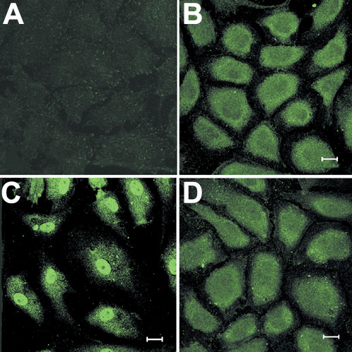
Figure 6. Localization of Cdk4 in response to FGF-2 stimulation
When CEC reached approximately 80% confluency, they were treated with FGF-2 (10 ng/ml) in the presence or absence of anti-PLC-g1 antibody for 24 h. Cells were then fixed and stained for Cdk4. A: CEC maintained in DMEM-10 and stained in the absence of anti-Cdk4 antibody. B: CEC maintained in DMEM-10 and deprived of serum for 24 h before staining for Cdk4. C: CEC treated with FGF-2 (10 ng/ml) for 24 h. D: CEC simultaneously treated with FGF-2 and anti-PLC-g1 antibody (5 ml) for 24 h. These images are representative of all three experiments. Bar is 20 mM.