![]() Figure 5 of
Lee, Mol Vis 2002;
8:17-25.
Figure 5 of
Lee, Mol Vis 2002;
8:17-25.
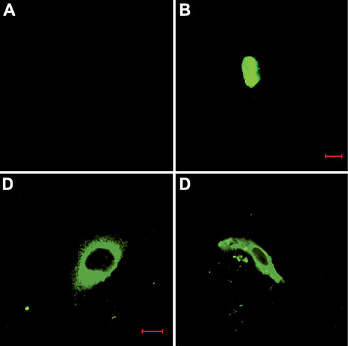
Figure 5. Localization of endocytosed anti-PLC antibodies
CEC were simultaneously treated with FGF-2 (10 ng/ml) and one of the anti-PLC antibodies for 24 h. Cells were then fixed and stained with FITC-conjugated secondary antibody as described in the text. A: CEC stained with FITC-conjugated secondary antibody. B: CEC treated with exogenous anti-PLC-b1 antibody for 24 h before staining with the secondary antibody. C: CEC treated with exogenous anti-PLC-g1 antibody for 24 h before staining with the secondary antibody. D: CEC treated with exogenous anti-PLC-d1 antibody for 24 h before staining with the secondary antibody. These images are representative of all three experiments. Bar is 10 mm.