![]() Figure 6 of
Boyle, Mol Vis 2002;
8:226-234.
Figure 6 of
Boyle, Mol Vis 2002;
8:226-234.
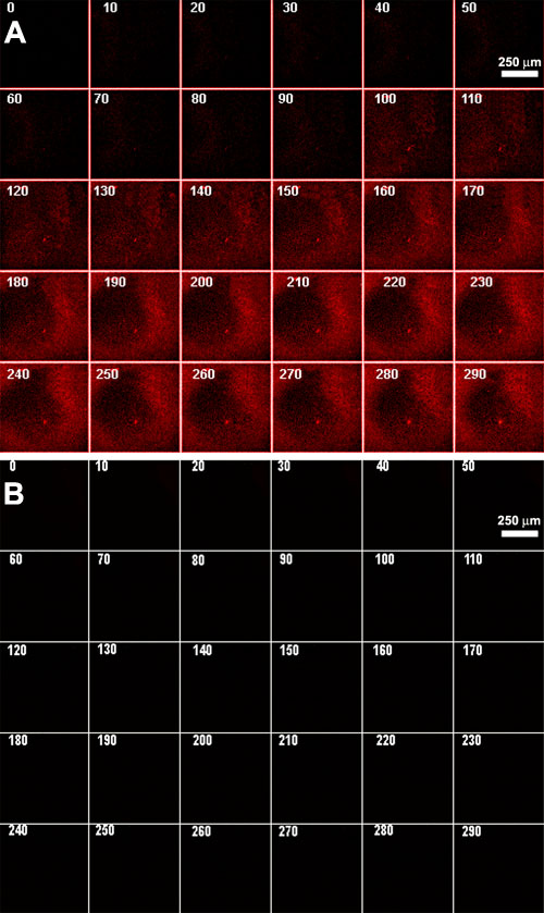
Figure 6. Real time analysis of DNA internalization
Real time fluorescence confocal microscopy of the epithelial cells in lenses incubated with fluorescently labeled plasmid DNA. Numbers accompanying each panel designate the time (min) of incubation with fluorescently labeled macromolecule. All the panels in A and all the panels in B correspond to the same field of epithelial cells. The darker regions of the fields in A and B correspond to regions of epithelial cells that are out of the plane of focus. All parameters used in the confocal microscopy were the same for A and B. A: Plasmid DNA without encapsulation. B: Plasmid DNA with prior encapsulation using GenePORTER liposomes.