![]() Figure 5 of
Boyle, Mol Vis 2002;
8:226-234.
Figure 5 of
Boyle, Mol Vis 2002;
8:226-234.
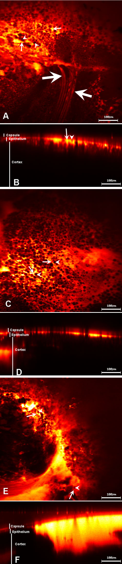
Figure 5. Confocal microscopy of dextran and other crystallins
Confocal microscopy of lenses incubated with fluorescently labeled β-crystallins, γ-crystallins, and dextran, after 290 min of incubation. A: The anterior epithelial cell layer is shown in the plane of focus. Fluorescently labeled γ-crystallins were preferentially seen in the cytoplasm (arrows) of epithelial cells and a few underlying fiber cells (large arrows), rather than in the nucleus (arrowheads). B: A z-scan of the lens in A. C: The anterior epithelial cell layer in the plane of focus showed the same localization (as seen in A) with fluorescently labeled β-crystallins. D: A z-scan of the lens in C. E: Preferential localization of fluorescently labeled dextran in the cytoplasm (arrowheads) and not in the nucleus (arrows) of the focal plane containing epithelium. F: A z-scan of the lens in E.