![]() Figure 1 of
Boyle, Mol Vis 2002;
8:226-234.
Figure 1 of
Boyle, Mol Vis 2002;
8:226-234.
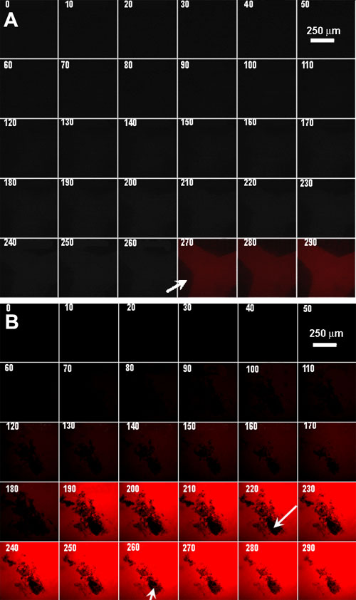
Figure 1. Real time analysis of α-crystallin internalization
Real time fluorescence confocal microscopy of epithelial cells in intact lenses incubated with fluorescently labeled α-crystallins. Numbers accompanying each panel designate the time (in min) of incubation with fluorescently labeled macromolecule. All panels in A, and all panels in B correspond to the same field of epithelial cells. The darker parts of the fields in A and B (arrows) correspond to regions of the epithelium that are out of the plane of focus. All parameters used in the confocal microscopy were the same for A and B. A: α-crystallins without encapsulation. B: α-crystallins with prior encapsulation using BioPORTER liposomes.