![]() Figure 4 of
Wistow, Mol Vis 2002;
8:205-220.
Figure 4 of
Wistow, Mol Vis 2002;
8:205-220.
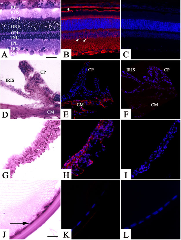
Figure 4. Localization of oculospanin in the rat eye by confocal immunofluorescence
A rabbit polyclonal antibody generated against an oculospanin peptide (OCSPp1) was used to examine the distribution of this protein in ocular tissues. The figure shows hematoxylin and eosin stained cryosections of retina (A), ciliary body (D), iris (G) and lens (J). Panels B, E, H, and K show corresponding sections with OCSPp1 immunolabeling. Primary antibody was omitted from negative controls (C, F, I, L) for each of the tissues. In the retina, oculospanin immunolabeling is prominent in the nerve fiber layer (NFL), ganglion cell layer (GCL), inner plexiform layer (IPL), outer limiting membrane (OLM), and the retinal pigment epithelium (RPE). A small population of retinal neurons located at the inner margin of the inner nuclear layer (INL) is immunolabeled (arrowhead). Separation of the rod outer segments (ROS) and apical surface of the RPE (*) is an artifact of tissue processing. In the ciliary body (E), immunolabeling is present in the ciliary muscle (CM) but not the ciliary processes (CP). Oculospanin immunolabeling was observed in the iris (H) but not the epithelia (arrow) or fiber cells of the lens (K). In panels A-I, the bar represents 40 μm; in panels J-L the bar represents 20 μm.